Alexandra Nikitas
Announcing the 2021 CRF Young Investigator Awards

The Cancer Research Foundation is pleased to announce the 2021 Young Investigator Awards
The Cancer Research Foundation just named six new CRF Young Investigators and we are thrilled to introduce them. The CRF Young Investigator Award is our predominant grant making program. It was established nearly 30 years ago to address a common problem in the early part of a cancer researcher’s career: to secure funding as a principal investigator, a scientist must present primary data, but to gather that data a scientist must have funding. The Cancer Research Foundation Young Investigator Award provides just that – each researcher receives $100,000 to gather primary data and establish themselves as a principal investigator.
These awards also allow the CRF to support and promote the belief that fighting cancer requires the best new ideas from a variety of different scientific fields, even those that are not traditionally cancer focused. While oncologists and pathologists on the front lines continue to lead the fight against cancer, relatively new contributions from fields such as data science, molecular chemistry and the study of genomics, proteomics and epigenetics are pioneering in cancer science and need to be supported.
Diversity and breadth of scientific research is a hallmark of our grants. Among the 2021 Young Investigators, we have scientists working in artificial intelligence and molecular engineering. We have researchers working on new models built from engineered human stem cells and scientists looking for ways that we might leverage drugs that we already know are safe. We have researchers looking at harnessing the mechanisms of DNA repair and immune system recruitment to kill cancer cells and stop their growth. We have two different projects concentrated on glioblastoma, focused on completely different cancer system problems, hoping to better understand this deadly disease. The Cancer Research Foundation believes that this diversity in different fields of research and schools of thought allows us to make the most impact in cancer science.
The diversity of ideas and science needed to extend cancer knowledge also requires that we support diversity among the researchers we fund. The Cancer Research Foundation is dedicated to the idea that we can only answer the wide variety of ways that cancer impacts different people with an equally diverse group of the best and brightest scientific minds. Our last few years of grants have been informed by our desire to support women and minorities in STEM and to reflect our belief that the scientists we fund should mirror all the different people affected by cancer. This year’s Young Investigators reflect our values and our commitment to diversity.
Meet the 2021 CRF Young Investigators
Click on a Young Investigator’s name to learn more
Huanhuan Joyce Chen, PharmD, PhD – Assistant Professor – University of Chicago
“Detecting and Decoding Polarization of Tumor-Associated Macrophage Using Human Pluripotent Stem Cell-Derived Cellular Sensors”
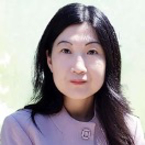
Dr. Chen proposes a new model in which the macrophages and cancerous cells are generated from the same cell source of human pluripotent stem cells (hPSCs), including human embryonic stem cells and inducible pluripotent stem cells. To examine the behaviors of tumor associated macrophages (TAMs) with greater precision, she will further engineer a genetic sensor in the TAMs that enables time-lapse detecting and capturing of the cells undergoing a “pro-tumor” or “anti-tumor” functional switch during disease progression.
Peiwen Chen, PhD – Assistant Professor – Northwestern University

“Targeting Macrophages and Microglia in PTEN-deficient GBM”
Dr. Chen intends to study a type of Glioblastoma (GBM) that is deficient in PTEN, a type of protein that controls cell division and acts as a tumor suppressor. He is particularly interested in the way that the cytokines LOX and OLFML3 allow the infiltration of Tumor Associated Macrophages and Microglia. Inhibiting these cytokines might be a way to allow immunotherapies to work in GBM.
Abby Green, MD – Assistant Professor – Washington University in St. Louis
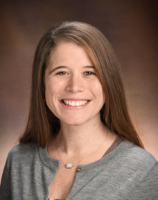
“The Double-edged Sword of APOBEC3 Mutagenesis in Cancer”
Dr. Green proposes a study of the APOBEC3 family of proteins. At some levels these proteins can cause cell death and could be used to combat cancer. However, at lower levels APOBEC3 proteins can create DNA mutations that actually lead to cancer. Using a unique chicken cell-based model, Dr. Green hopes to understand how the APOBEC3 proteins are related to cancer development.
Xavier M. Keutgen, MD, FACS – Assistant Professor – University of Chicago

“The role of MEN1 loss of function and Estrogen Receptor α inhibition in radiosensitizing Pancreatic Neuroendocrine Tumors”
Dr. Keutgen’s work is focused on a recent discovery that mutations in the MEN1 gene affect the DNA repair mechanism required by cancer cells to withstand radiation therapy. His hypothesis is that the MEN1 protein works together with the estrogen receptor (ESR1) protein to make cells radio resistant and that ESR1 inhibitors already being used in breast cancer treatment might be an effective additional therapy to radiosensitize pancreatic neuroendocrine tumors.
Allegra A. Petti, PhD – Assistant Professor – Washington University in St. Louis
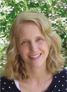
“ A Dynamic Molecular Atlas of Glioblastoma During Tumor Formation and Treatment Response”
Dr. Petti, a specialist in systems biology and genomic inquiry, plans to focus on the area at the outside edges of Glioblastoma (GBM) tumors and study differences in tumor organization from the center of a tumor to its margins. She hopes to better understand the relationships, both genomic and spatial, between tumor cells and immune cells in GBM using new technologies.
Samantha J. Riesenfeld, PhD – Assistant Professor – University of Chicago
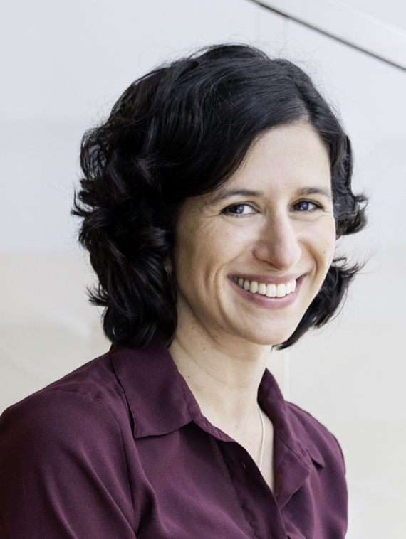
“Extracting Latent Transcriptional and Multi-omic Features from Digital Pathology to Guide Cancer Treatment“
Dr. Riesenfeld proposes to further the quest to use machine learning to identify key patterns in images of microscopic tissue structure, extending the use of such images from classifying cancer to learning “multi-omic” indicators that integrate information across DNA, gene expression, and chromatin structure. The goal is to build systems that, by leveraging limited, expensive molecular data, can powerfully use routine imaging to improve diagnostic and treatment decisions.


